Abstract
Clinical imaging is key to dental diagnostics and treatment planning, but traditional radiographs and 2D panoramic images have limitations. 3D Cone Beam Computed Tomography (CBCT) literally brings a new dimension to dentistry and its benefits enhance a wide range of clinical situations.
Learning Objectives
- Understand the benefits of cone beam computed tomography (CBCT)
- Learn about its clinical uses
- Know the areas for which it provides critical information
Cone Beam Computed Tomography (CBCT) imaging is transforming the way dentists acquire diagnostic information. CBCT scans provide greater imaging accuracy and detail for better diagnosis and treatment planning, improved execution of surgical procedures, and more information for assessing pathology and outcomes.1 Traditional full-mouth radiographic series and panoramic images deliver the basics for presurgical assessments, but in many cases, they lack the detail necessary to plan and execute treatment that will yield superior surgical outcomes.
CBCT is commonly used to:
- Extract teeth, including third molars
- Diagnose temporomandibular joint disorders
- Accurately place implants and fabricate surgical guides
- Locate pathology in the jaw
- Plan and assess root canal therapy and retreatment
- Visualize and diagnose root fractures
- Assess airways
- Perform cephalometric analysis and determine root location
As its cost has diminished, CBCT imaging has become more commonplace. When selecting CBCT equipment, be sure to understand what your present and future needs are, so you can select the proper field of view (FOV). Generally, as FOV increases, so does the cost of the machine.
Once you start using CBCT, you begin to realize when the information it provides is invaluable for planning purposes. There is never an instance in which I would find less information beneficial. But there are many cases for which routine 2D imaging is sufficient, and I want to follow the ALARA principles for patient exposure to radiation. When I decide that I need 3D imaging, it is because to plan the best treatment, I need the additional details it offers. In addition, CBCT data is also important for other clinical decisions, such as determining whether a case is beyond my training and a referral for specialty care is necessary.
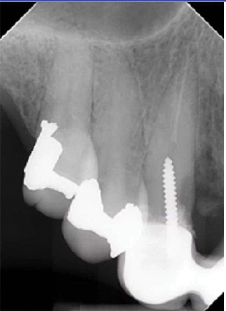
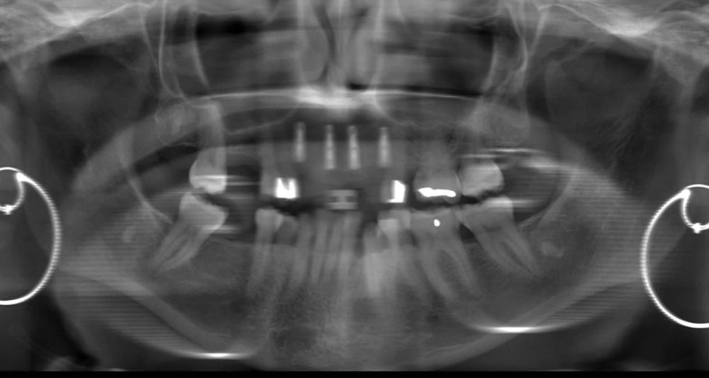
For me, the most important benefit of CBCT 3D imaging is determining whether an implant can be optimally placed or whether other treatment options should be explored. Routine 2D imaging (Figure 1) is always a good starting point, but when dealing with esthetic cases with multiple implants, CBCT 3D images makes treatment easier.
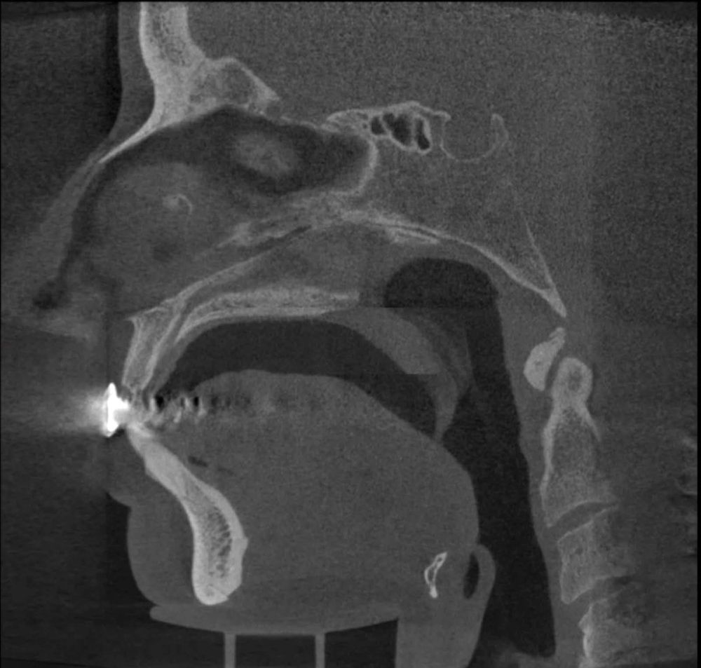
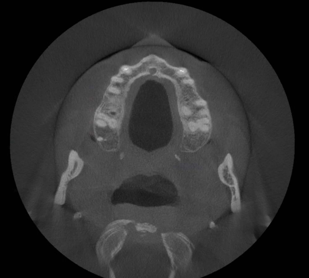
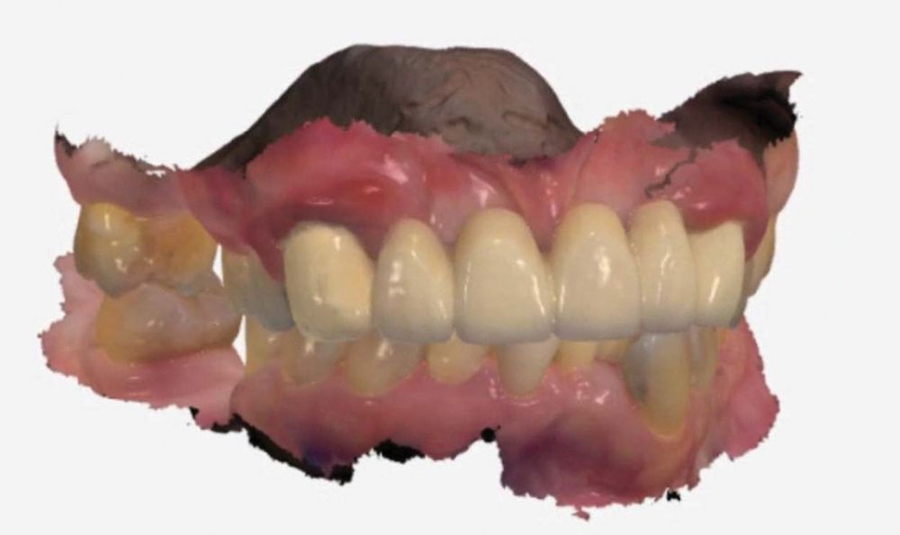
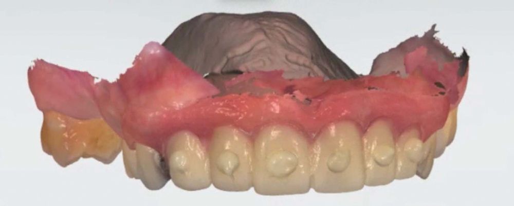
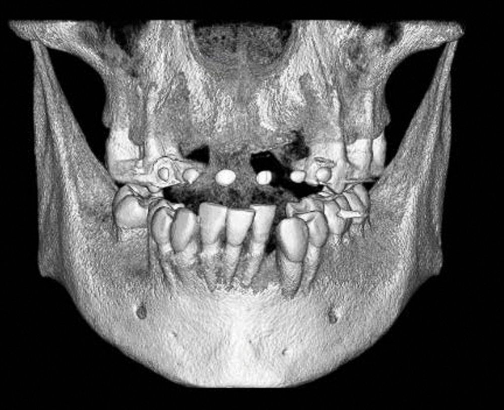
The bridge shown in Figure 1 was failing, and the patient requested an implant. A PreXion CBCT scan was obtained in my practice to determine the placement and the number of implants that were possible (Figures 2a, 2b). An intraoral scan was obtained for the fabrication of a temporary printed Valplast removable partial denture (RPD) (Figure 3).
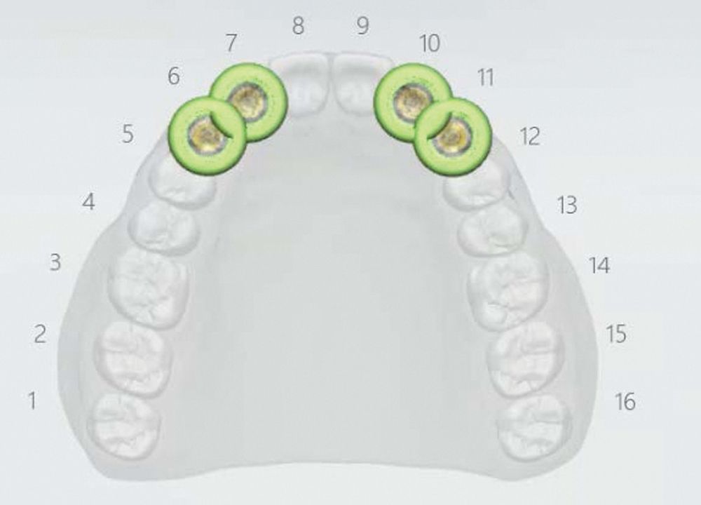
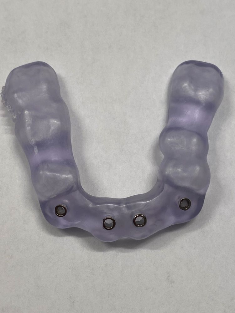
The teeth were then extracted and the RPD placed. After sufficient healing, the RPD was used as a guide by placing flowable composite on the front teeth. The RPD was placed in the mouth again, and the CBCT scan was taken for additional planning (Figures 4a, 4b). Merging of the 3D images allowed for proper implant planning and the printing of a surgical guide (Figures 5,6). The guide was inserted in the mouth, and the implants placed (Figure 7).
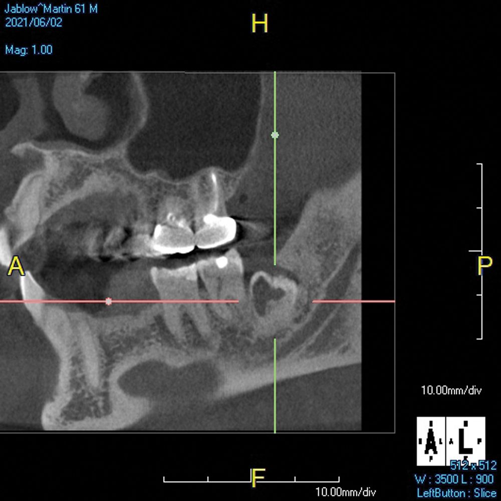
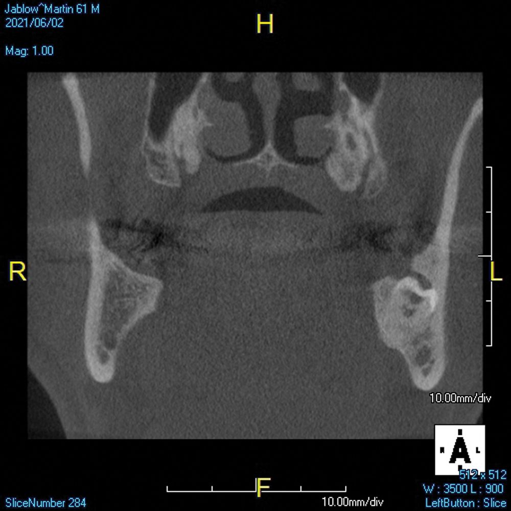
Impacted third molars are another scenario in which CBCT imaging gives you necessary data.2 Knowing how close the teeth are to the inferior alveolar nerve can make a better surgical approach possible and hopefully minimize trauma to the area and reduce the likelihood of a paresthesia (Figures 8a, 8b).
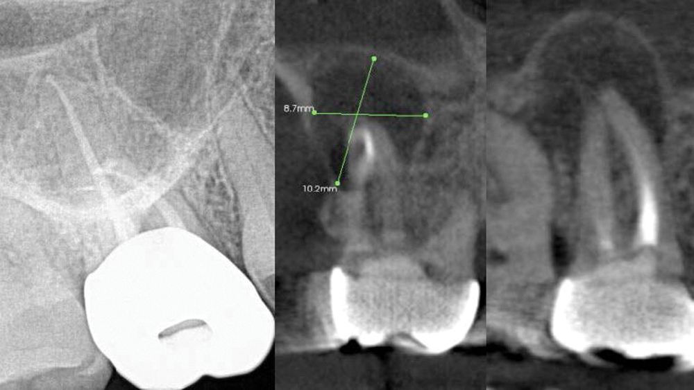
Endodontic cases can be challenging, especially those involving maxillary molars whose roots overlap into the sinus. CBCT imaging simplifies these cases, as the number of canals and the extent of pathology can be determined before initiating treatment (Figure 9).3 After viewing the CBCT images, it may be evident that the tooth is not restorable because of the extent of the lesion. It is rarely possible to determine this prior to surgery using only 2D imaging.
3D CBCT imaging captures more information about the patient’s anatomy and can reveal numerous health issues.4 I like to say that you never know what you may find while viewing a CBCT scan.
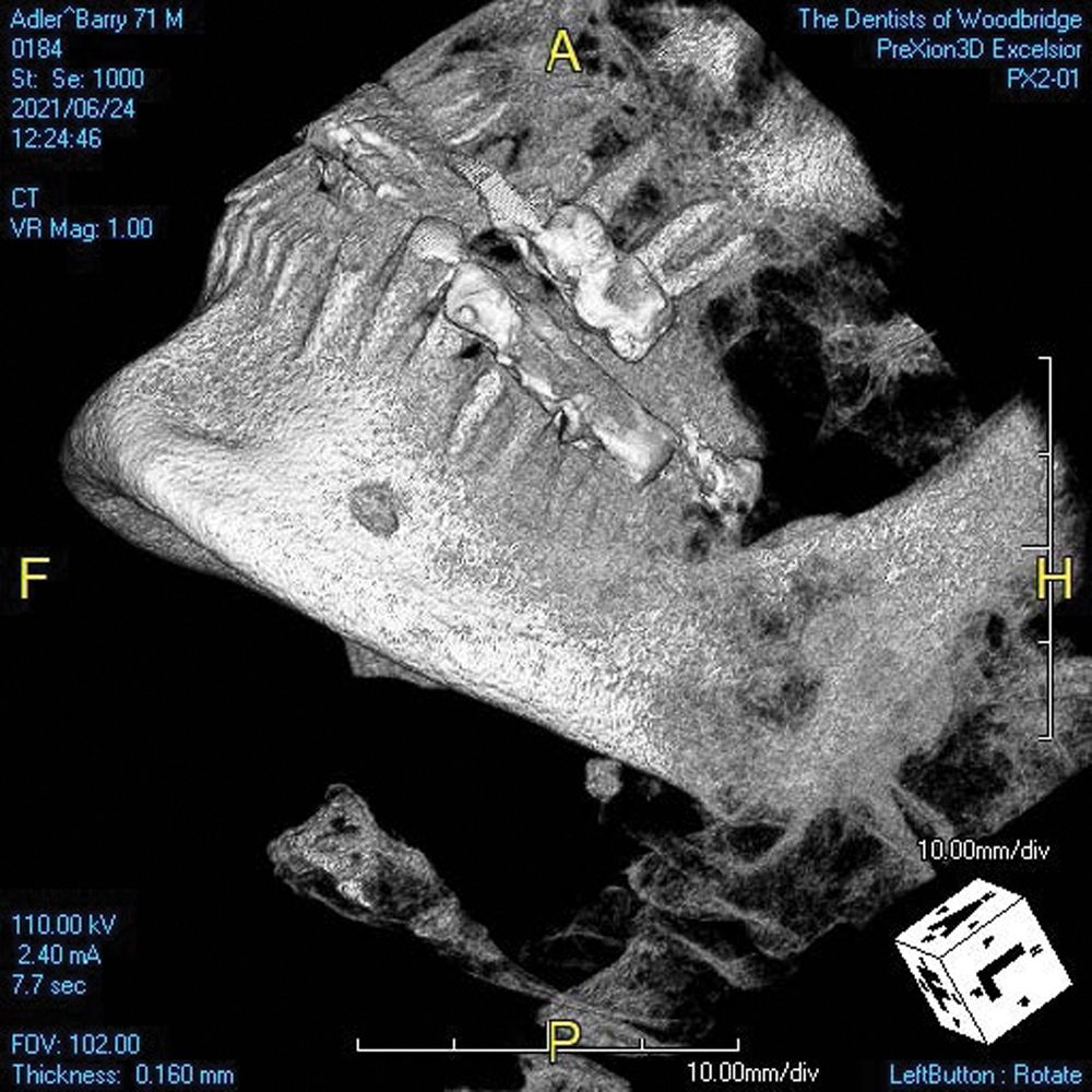
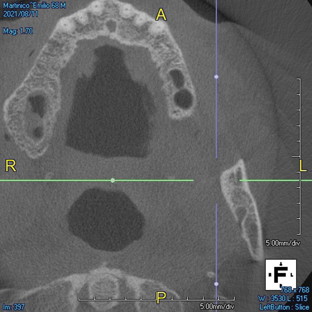
Here are 2 other findings I didn’t anticipate. Figure 10 shows a lymph node calcification. Figure 11 is from a patient who presented with pain, and pathology was found in the sinus, along with perforation of the buccal plate.
As you can see, these patients walk through your door every week. A CBCT scan takes the guesswork out of diagnosis and treatment planning. It can also help patients better understand treatment recommendations. We can show them the details and demonstrate why something is necessary or why a requested treatment won’t be possible. With benefits to clinicians and patients alike, it’s clear CBCT imaging plays a key role in the present and future of dentistry.
References
- Shukla S, Chug A, Afrashtehfar KI. Role of cone beam computed tomography in diagnosis and treatment planning in dentistry: an update J Int Soc Prev Community Dent. 2017;7(Suppl 3):S125-S136. doi:10.4103/jispcd.JISPCD_516_16
- Hermann L, Wenzel A, Schropp L, Matzen LH. Impact of CBCT on treatment decision related to surgical removal of impacted maxillary third molars: does CBCT change the surgical approach? Dentomaxillofac Radiol. 2019;48(8):20190209. doi:10.1259/dmfr.20190209
- Patel S, Brown J, Pimentel T, Kelly RD, Abella F, Durack C. Cone beam computed tomography in Endodontics — a review of the literature. Int Endod J. 2019;52(8):1138-1152. doi:10.1111/iej.13115
- Dief S, Veitz-Keenan A, Amintavakoli N, McGowan R. A systematic review on incidental findings in cone beam computed tomography (CBCT) scans. Dentomaxillofac Radiol 2019;48(7):20180396. doi:10.1259/dmfr.20180396

 Download Issue: Dental Products Report February 2023

