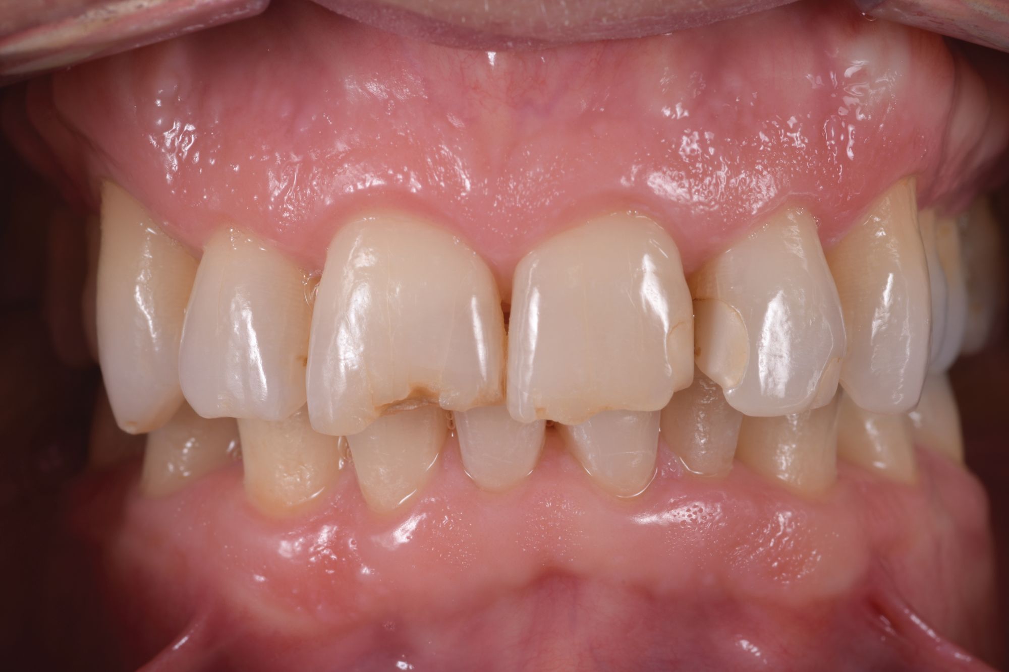
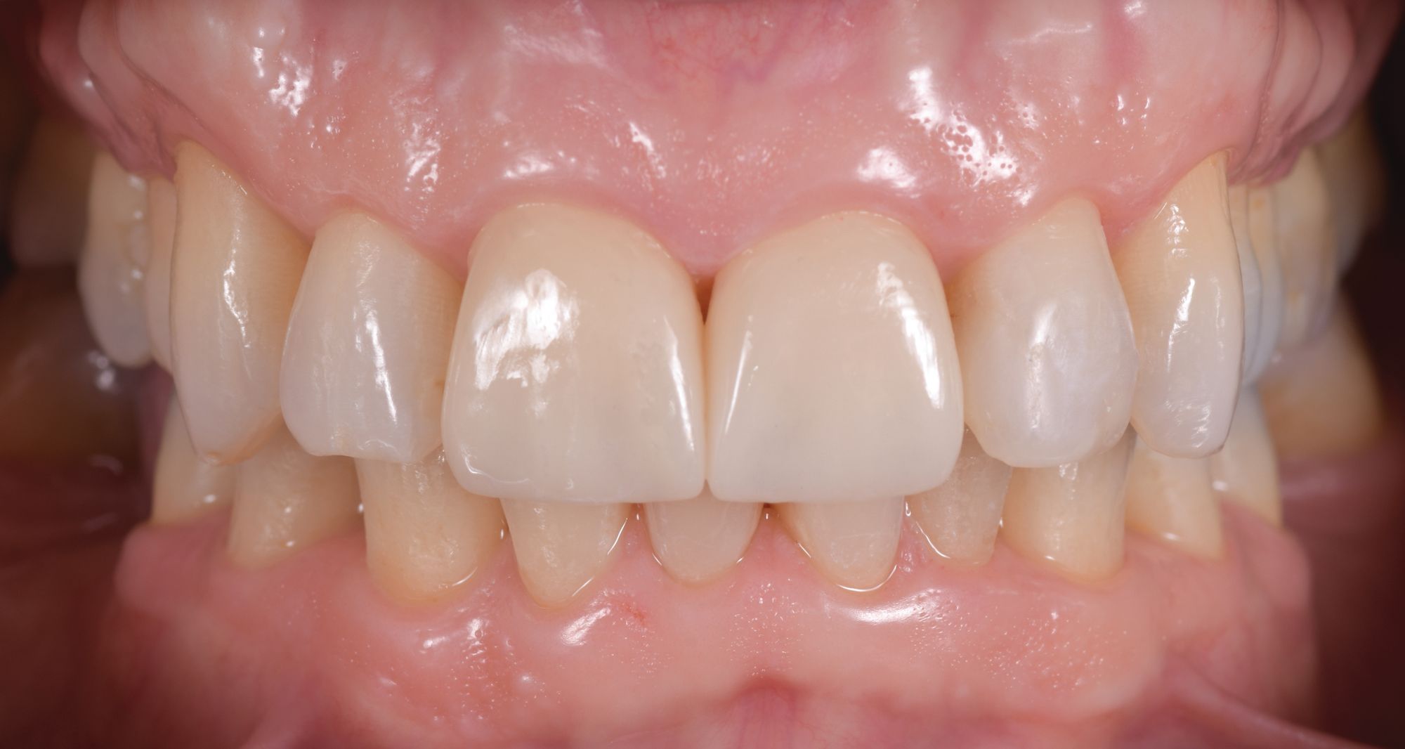
An intraoral image illustrating the condition of the maxillary incisors was taken with the CEREC Primescan (Dentsply Sirona) for patient education. The clinicalimage was shown to the patient, and upon being advised of the findings, he agreed to proceed with full contour crowns to restore both maxillary central incisors and a direct resin restoration for his maxillary left lateral incisor.
Clinical Objective
The clinical objective of this case was to restore the esthetic form and function of the maxillary incisors in the most predictable manner, utilizing the most appropriate materials and techniques for long-term results.
Technique
At the initial visit, a series of digital diagnostic photos was taken for smile design planning. Dual full arch digital impressions with the CEREC Primescan were taken and sent via CEREC Connect to the dental lab technician for the fabrication of a digital diagnostic wax-up of the maxillary central incisors.
At the time of the procedure, both failing composite restorations were removed and the recurrent caries excavated. CARIES DETECTOR (Kuraray America, Inc) was applied to assist in verification of complete caries removal. A composite buildup was placed on tooth #8 using CLEARFIL™ SE Protect, CLEARFIL MAJESTY Flow, and CLEARFIL AP-X composite resin (all Kuraray). A direct resin restoration was placed on #10 using CLEARFIL SE Protect Primer & Bond, MAJESTY Flow, and MAJESTY composite resin.
CLEARFIL SE Protect provides excellent bond strength and has an antibacterial cleansing effect. The author has also found that patients report no incidence of postoperative sensitivity with cases utilizing CLEARFIL SE Protect.
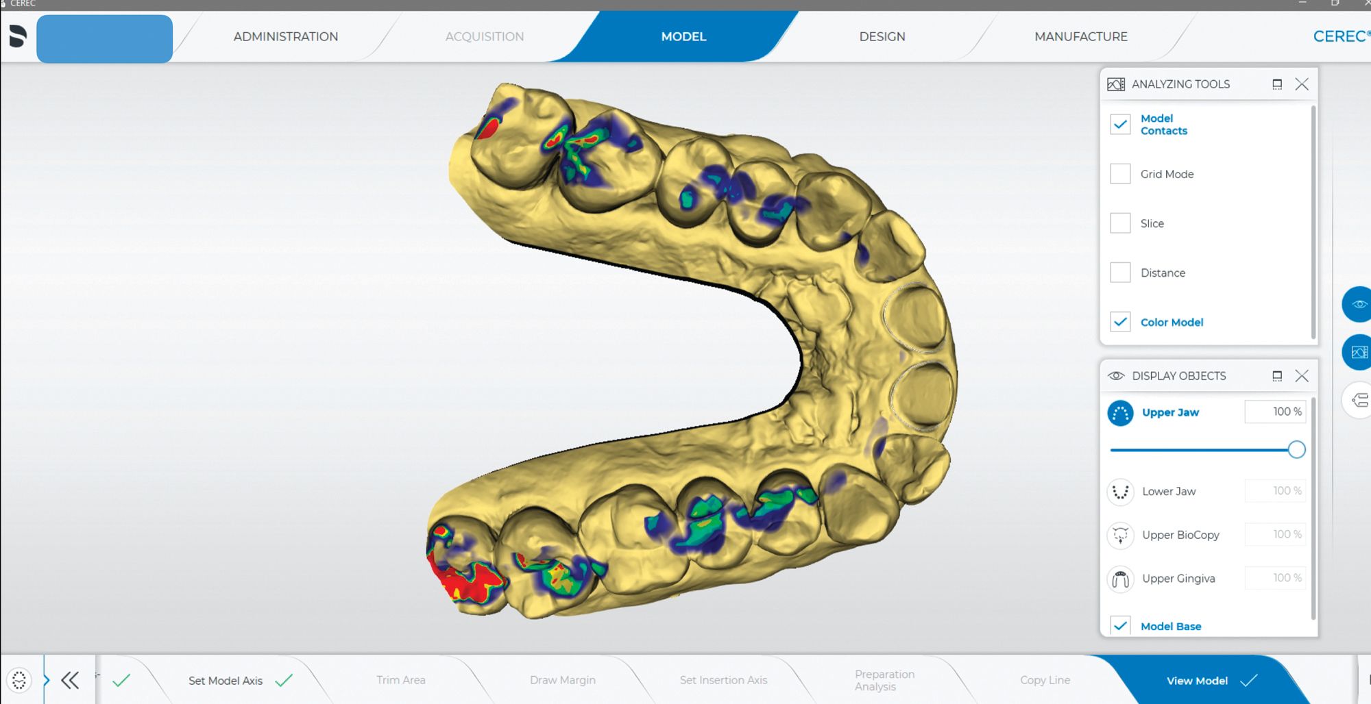
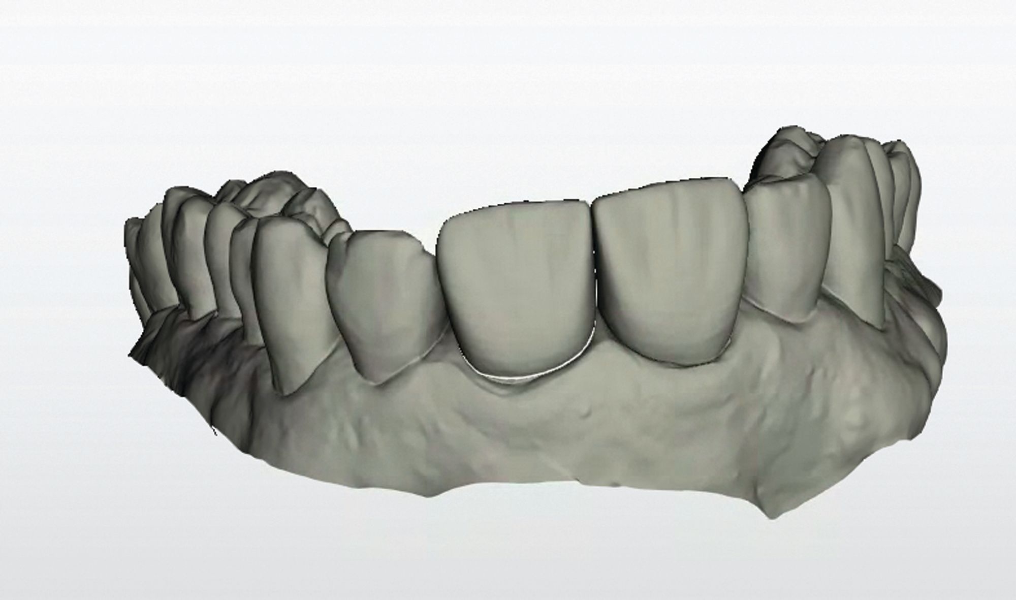
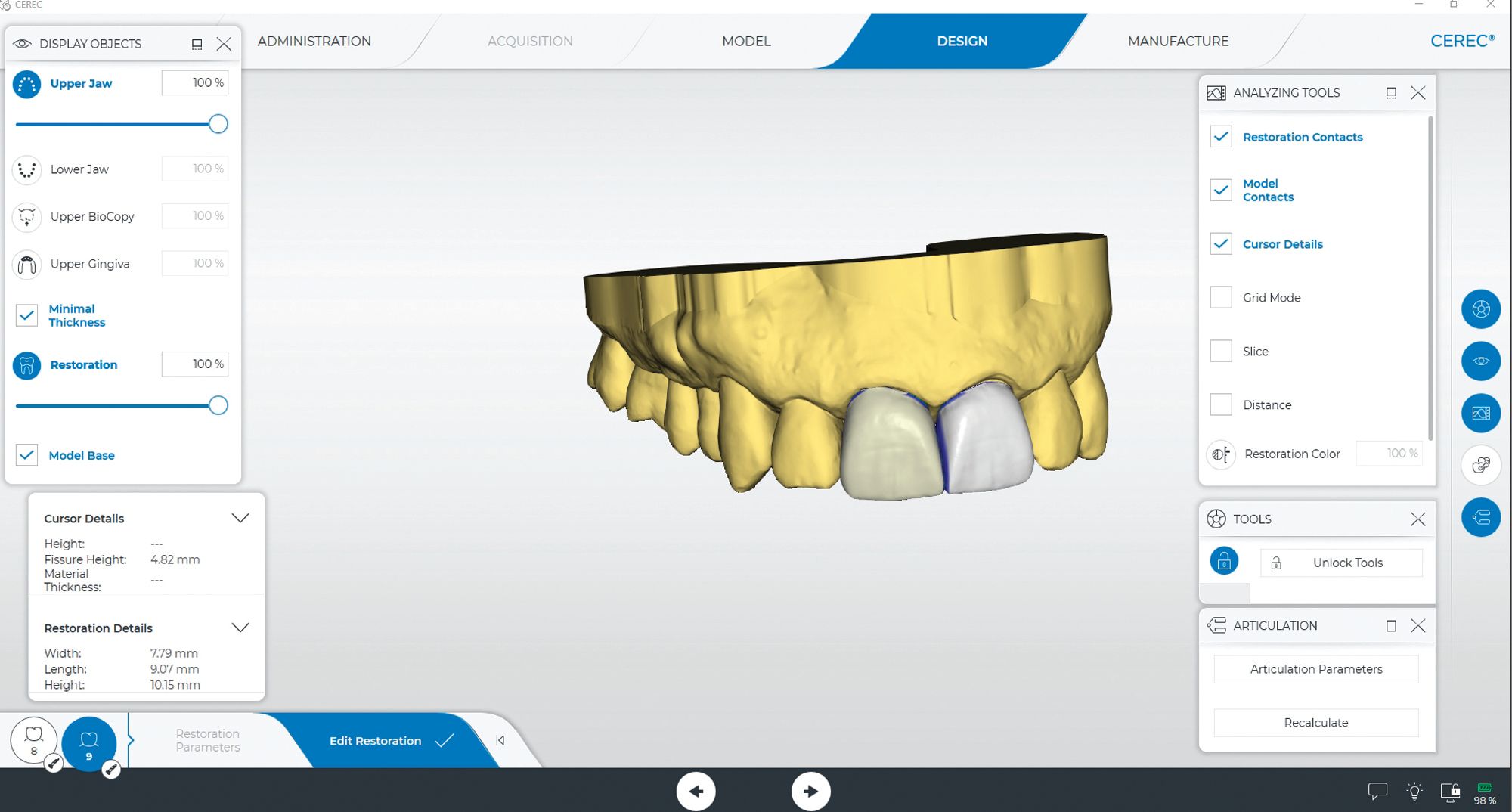
After the composite buildup and minimal preparations with conservative chamfer margins, preparations were completed. The lower jaw, upper jaw, and buccal bite were recorded using the CEREC Primescan in the acquisition phase. The virtual models were then created (Figure 2).

 Download Issue: Dental Products Report June 2023