Focusing on Ideal Digital X-Rays
DentiMax IT Specialist Micah Huish explains how he sets up dental practices for imaging success, ensuring their radiographs look exactly the way they prefer.
DENTAL PRODUCTS REPORT®: Can you tell us what you do for dental practices?
Micah Huish: We set [them] up with our software and sensor [DentiMax Imaging Software and Dream Sensor], or we go in with our sensor into their software, get them working with it, see how it functions, [and] make sure they’re familiar and comfortable with all that.
The fun part comes [when] we do our dial-in process. We have [them] take a couple [of] test x-rays on actual teeth. Usually, we recommend doing [them] on an employee; that way, we have time to talk and make changes. Then we find out what [they] want: [their] perfect-looking image. And then we go in with precapture filters and adjust those filters so that it’s the best diagnostic image that we can get for [them].
DPR: Can you tell me about some of the settings and controls you have to adjust the x-rays?
MH: In most cases, all software [can] adjust the image. After you take the x-ray, it has some type of filter: brightness/contrast tool, gamma tool, [or] something like that. We [can] adjust the capture filters with our sensor and software so that the image appears more [as] you want it right off the bat.
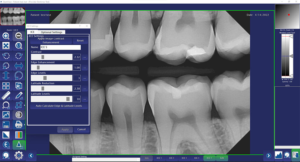
With our software we can adjust those same filters. We can adjust the gamma, sharpen, brightness/contrast and more. We can even adjust the level of contrast or the type of sharpen depending on how you like seeing your image. Having the ability to adjust all of these filters both pre and post capture is great because one practice my like a more black and white super sharp image whereas another might like a more mono-gray image that’s not as sharp. With our sensors, tools, and filters we have the ability to give practices and doctors the image they prefer. Another huge benefit is that we can customize the settings in each room. This is perfect for those practices that may have more than one provider. Being able to adjust the settings allows for all the providers to get an image they like seeing rather than having to see just what’s already there.
We have normalized filters, we have unsharpened filters; there’s a whole bunch of different filters we can adjust to make the image [appear] more [as] you want it.
DPR: Is there a typical x-ray look most practices are looking for?
MH: That’s the thing I think is amazing. You can go and work with 6 doctors in [a] day. And I’ll get [an image and think] that’s the image they’re going to love because [it] looks just like the one the last doctor loved. And [the doctor will say], “No, I don’t want anything like that. I want it to be completely different.”
Most doctors tend to like the image they’re used to, what they’ve had in the past, what they’ve used for the [past] few years. So we go in and adjust based on that. But no 2 doctors like the same image. There may be some similarities, but you can show 10 doctors the same image: 5 of them will say it’s amazing, [and] 5 of them will say it’s horrible.
DPR: If they don’t all agree on the type of image they like, is there an image you know is universally what they don’t want?
MH: We probably hear most that they don’t want a grainy image. They feel it adds in things that aren’t there. It makes it harder to see interproximal caries or decay and any other issue the teeth may have.
The second-most common thing they want to have is a good level of contrast, so they can distinguish the DEJ [dentinoenamel junction]. They can see the apex of the root very clearly, they can see caries very easily.
So those are the 2 things they want most: not to have a grainy image and to have enough contrast, because if they don’t have enough, it’s too hard to distinguish between the areas of the teeth.
DPR: Are there controls in the practice they can adjust themselves, or is it something they should set up initially and then continue to shoot with the same settings every time?
MH: [For] the settings we adjust, usually once we have them set up, you just keep moving forward [and] you don’t look back; you’ll just continue getting the same good image as long as [practice staff are] being consistent.
Exposure time plays a big factor, and the technique of the staff, plus how far away [or] close to the face, [whether] they’re lining up their shots properly. All that goes into what causes the image to look good or bad.
When they slow down and take their time, they should get a consistent image with the settings we lock in, once we get them dialed in.
DPR: What does that dial-in process look like?
MH: Again, that can depend on the doctor. Different specialties like different features on an image. An endodontist [is] going to like a sharp image in most cases, because that allows them to see the apex and all the areas of the teeth they specialize in.
When you start taking x-rays, you get used to reading a specific style or look of x-rays. When you switch to a new sensor, you want it to be better, similar to what you’re used to, or a mix between the 2. Doctors just want a great image that is familiar to them, better than what they have and/or easier to diagnose with. With our sensor, we do this daily.
DPR: If someone adds a DentiMax sensor to their practice not as a replacement but as an extra sensor, are you able to dial in that sensor to mimic the other sensor the practice is using?
MH: All sensors take different images. They have different components, products, filters, things like that. But we have made integrations that allow [them] to be very, very close. [Many] doctors can’t tell the difference between the 2 images, but we can usually tell the difference.
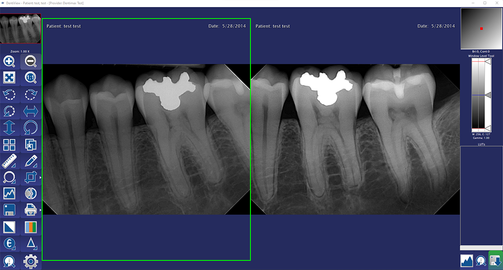
Most software automatically applies a filter after you capture. If you were to look at [an] x-ray with the sensor and what it captured before [the] filter was added, you’d probably be surprised at [the x-ray] before [the] filter. Most offices use filters; a lot of filters make the image, to my eye, much more pleasant [and] readable.
Again, that is a complete preference based on what you’re used to. But I recommend that doctors play with filters [and] see what helps them diagnose better. Because the more they’re diagnosing, the better they’re going to do.
DPR: Are there specific filters you find doctors [use] more than others?
MH: The one we see most often is the brightness/contrast filter. Hygienists use it to adjust the image to help diagnose. Doctors use it to adjust the image to diagnose. It changes how light or dark the image is and add more or less contrast to help see the different parts of the teeth which can help with seeing things such as interproximal caries.
It also helps if you’re not sure [about] something. A lot of times caries will stay a darker color no matter [how] you adjust that brightness/contrast tool. It helps reiterate that caries [are] right there.
DPR: Can you see different things in a darker or lighter x-ray image?
MH: From my experience, hygienists usually like the image a little lighter, doctors usually like them a little bit darker, [with] some variation.
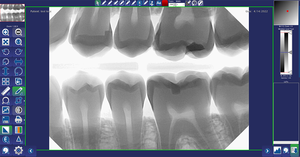
But the problem with an image that is too dark is that it can introduce the effects of burnout even if the image isn’t overexposed. Super dark images start to almost eat away part of the image causing areas of the tooth or teeth to not be visible in the image. You may not be able to see caries because the image is so dark that it’s hidden behind some of that shadowing. So those are the downfalls of going too dark.
But when you’re darker, your pulp chambers usually pop more, allowing you to see the apex easier. You can see the apex of the root visibly because the image is darker.
When they’re too light, you might not see caries because it’s so washed out that there are no distinguishing features of the tooth, [and] you can’t see the DEJ clearly. You can’t determine what the different colors of each part of the tooth are.
DPR: How does communication with a practice go? I imagine most doctors don’t know how to ask for what they want in an image, so how do you work with them to get that image?
MH: Sometimes it’s a guessing game. We’ll hear the word sharpen, and my definition of a sharper image would mean the lines are crisp, they’re sharp, they’re hard edge. But somebody else may be using the word sharp, [and] they want more of a contrast: more colors, brighter colors, more defined colors, things like that. But we usually ask questions.
We usually start the call by having them show us what they like [and] what they want to see. And that allows us to try and understand some of their verbiage or terminology a little better, so when we [ask] how they want their image, it makes it easier for us to understand them. And if we don’t understand, we’ll adjust.
We [can] show them what we think they mean. And if that’s not [it], we can undo and change to whatever they do mean. We like to have them show us what they like or dislike. In some cases, it didn’t have anything they may like, and tell us why they like it. And then we’ll do our best to either mimic that image as close as possible or [get] something better. I love to see how much better I can make their image.

 Download Issue: Dental Products Report August 2022